


AOKWIT Outdoor Knowledge Course – Outdoor First Aid Knowledge (Part 2)
VI. How to Handle Acute Abdominal Pain?
Sudden, severe, and rapidly changing abdominal pain is termed acute abdominal pain. The causes of acute abdominal pain can be classified into three categories:
- Acute intra-abdominal organ diseases (e.g., appendicitis, perforated ulcers).
2. Thoracic diseases causing referred abdominal pain (e.g., pneumonia, myocardial infarction).
3. Systemic diseases (e.g., metabolic disorders, allergic reactions, or toxin exposure) that irritate abdominal nerves. Among these, acute intra-abdominal organ diseases are the most common cause.
Characteristics of acute intra-abdominal organ-related pain:
- Localized pain: The pain location typically corresponds to the affected organ. For example:
- Upper-middle abdominal pain: Often indicates gastric or duodenal ulcer perforation.
- Lower-right abdominal pain: Suggests acute appendicitis.
2. Pain nature: Closely related to the underlying cause.
- Acute inflammation (e.g., cholecystitis): Pain develops gradually (over hours to days), starts as tenderness, and progresses to rebound tenderness (sharp pain upon sudden release of pressure) and abdominal muscle rigidity (hardened abdominal wall).
- Gastrointestinal perforation: Sudden, intense, knife-like pain that spreads rapidly, accompanied by generalized tenderness, rebound tenderness, and rigidity.
When to seek immediate medical attention:
1.Severe pain with sweating, inability to stand, or unrelieved pain after taking analgesics.
2.Pain causing confusion, pallor, cold sweats, bradycardia (slow pulse), or chills.
3.Board-like rigidity of the abdomen (indicative of peritonitis).
4.Persistent vomiting or inability to pass stool.
Pre-hospital care guidelines:
- Positioning:
- Let the patient rest in a quiet environment with loosened clothing.
- Allow the patient to adopt a comfortable position (e.g., lying on their side if vomiting).
2. Do NOT:
- Provide food, drinks, or unprescribed medications (especially analgesics, which may mask symptoms).
- Apply heat to the abdomen.
3. Monitor vital signs:
- Check for fever, respiratory rate, pulse, and blood pressure.
4. Symptom management:
- For vomiting: Place an ice pack on the stomach to reduce nausea (avoid suppressing vomiting).
Patient positioning for specific conditions:
- General cases: Lie flat with limbs extended.
- Hypertensive cerebral hemorrhage: Elevate the head slightly to reduce intracranial pressure.
- Unconscious patients: Turn the head sideways to prevent aspiration of vomit.
- Shock: Slightly lower the head to improve cerebral blood flow.
- Respiratory distress: Use a semi-seated position to ease breathing.
Transportation precautions:
- Gentle handling: Move the patient carefully to avoid exacerbating injuries.
2. Fracture management:
- Support fractured limbs to prevent movement.
- Stabilize spinal injuries by keeping the back straight.
- Immobilize head trauma to minimize brain injury.
3. Stretcher use: Maintain a horizontal position when ascending/descending stairs.
4. Monitor en route: Continuously check respiration, pulse, and consciousness.
5. Keep warm: Cover the patient with blankets or dry clothing if wet.
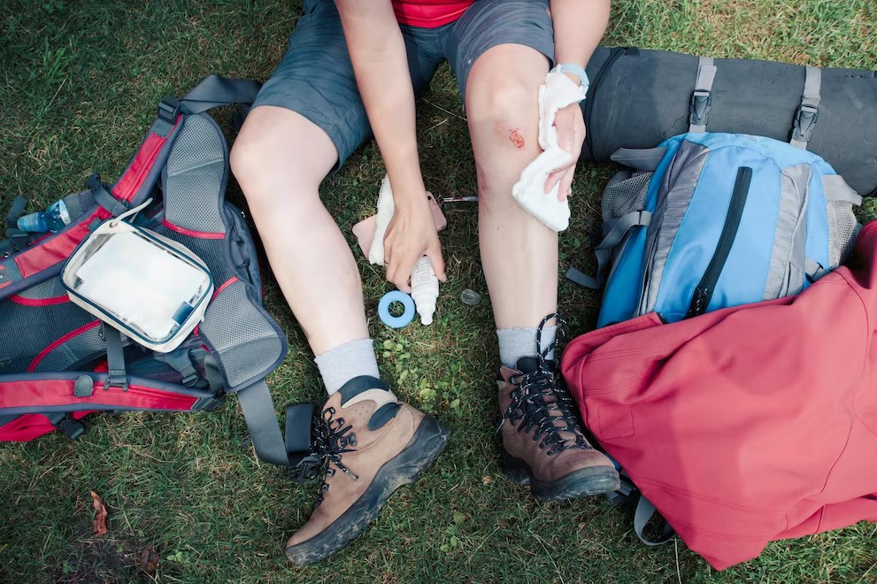
VII. Ankle Sprain Management
Causes: Typically due to sudden twists, missteps, or uneven landings. Symptoms include swelling, pain, bruising, and potential fractures.
Immediate first aid (RICE protocol):
- Rest: Avoid weight-bearing on the injured ankle.
2. Ice: Apply an ice pack wrapped in cloth for 15–20 minutes every 1–2 hours (prevents tissue damage).
3. Compression: Use an elastic bandage to reduce swelling (ensure it is snug but not restrictive).
4. Elevation: Keep the ankle elevated above heart level to minimize swelling.
Clinical assessment steps:
- Visual inspection: Check for deformity, swelling, or discoloration.
2. Palpation: Identify localized tenderness (e.g., lateral malleolus for lateral ligament injury).
3. Functional tests:
- Inversion test: Gently tilt the foot inward to assess lateral ligament integrity.
- Eversion test: Tilt the foot outward to evaluate medial ligaments.
- Anterior drawer test: Stabilize the lower leg while pulling the heel forward to check for excessive anterior movement (indicative of ligament tear).
Treatment based on severity:
- Grade I (mild): Continue RICE protocol; gradual return to activity in 1–2 weeks.
- Grade II (moderate): Immobilize with a brace; physical therapy for 3–6 weeks.
- Grade III (severe): Refer for imaging (X-ray/MRI) to rule out fractures; surgical consultation if needed.
Rehabilitation phases (7-stage recovery):
- Acute phase (0–72 hours): Focus on reducing swelling and pain (ice, elevation).
2. Subacute phase (3–7 days): Begin gentle range-of-motion exercises (e.g., ankle alphabets).
3. Early strengthening (1–2 weeks): Resistance band exercises for dorsiflexion, plantarflexion.
4. Moderate strengthening (2–4 weeks): Weight-bearing exercises (e.g., heel raises).
5. Advanced strengthening (4–6 weeks): Balance training (e.g., single-leg stands).
6. Sport-specific drills (6–8 weeks): Agility drills, lateral movements.
7. Full return (8+ weeks): Gradual reintroduction to sports with protective taping.
Advanced rehabilitation techniques:
- Contrast hydrotherapy: Alternate warm (38–40°C) and cold (10–16°C) water immersion to improve circulation.
- Proprioceptive training: Use wobble boards to restore joint stability.
- Gradual loading: Progress from partial to full weight-bearing with crutch support.
VIII. Managing Travel-Related Emergencies
- Fainting (Syncope):
- Lay the patient flat with legs elevated (improves cerebral blood flow).
- Acupressure: Press the Hegu acupoint (LI4, between thumb and index finger) firmly for 2–3 minutes.
- Loosen tight clothing; ensure ventilation.
2. Headache:
- Apply pressure to Taiyang acupoints (temple regions) using circular motions for 2–3 minutes.
- Hydrate and rest in a dark, quiet environment.
3. Stomachache (Gastralgia):
- Massage the Zusanli acupoint (ST36, 4 finger-widths below the knee cap) for 3–5 minutes.
- Avoid spicy or greasy foods; sip warm water.
4. Hypertensive Crisis:
- Press the Laogong acupoint (PC8, center of the palm) and fingertips alternately to lower blood pressure.
- Administer prescribed antihypertensives if available; seek urgent medical care.
5. Angina Pectoris:
- Apply pressure to the Jianjing acupoint (base of the middle fingernail) for 3–5 minutes.
- Administer sublingual nitroglycerin if prescribed; call emergency services.
6. Muscle Cramps:
- Pinch the Renzhong acupoint (GV26, philtrum groove) firmly for 20–30 seconds.
- Stretch the affected muscle gently; hydrate with electrolyte solutions.
7. Asthma Attack:
- Press and massage the Yuji acupoint (LU10, midpoint of the thumb’s metacarpal bone) for 3 minutes.
- Use a rescue inhaler (e.g., albuterol); maintain calm breathing.
8. Nosebleed (Epistaxis):
- Pinch the heel opposite the bleeding nostril (left nostril → right heel; right nostril → left heel).
- Lean forward slightly; apply ice to the bridge of the nose.
IX. Snakebite First Aid and Management
Symptoms by venom type:
- Neurotoxic venom (e.g., cobras): Systemic symptoms like ptosis, respiratory paralysis, blurred vision.
- Hemotoxic venom (e.g., vipers): Local necrosis, hemorrhagic blisters, coagulopathy.
- Mixed venom (e.g., rattlesnakes): Combined neurotoxic and hemotoxic effects.
Immediate actions:
- Stay calm: Minimize movement to slow venom spread.
2. Immobilize the limb: Use a splint or sling to keep the bite site below heart level.
3. Remove constrictive items: Rings, bracelets, or tight clothing near the bite.
4. Loose constriction band: Apply a lymphatic constriction band 5–10 cm above the bite (not a tourniquet). It should allow one finger underneath; loosen every 15–30 minutes.
Wound care:
- Clean the wound: Rinse with saline or clean water (avoid alcohol or ice).
2. Avoid incisions or suction: Modern guidelines discourage cutting or mouth suction due to infection risk.
3. Apply antivenom (if available): Follow manufacturer instructions (e.g., Nantong Jidesheng snake tablets).
Emergency evacuation:
- Transport the patient to a hospital with antivenom stocks immediately.
- Note the snake’s appearance (size, color, head shape) for identification.
Preventive measures:
- Wear snake-proof boots and gaiters in high-risk areas.
- Use a flashlight at night; avoid reaching into crevices.
- Carry a snakebite first aid kit with bandages and antiseptics.
X. Emergency Responses to Travel Illnesses
- Syncope (Fainting):
- Position: Supine with legs elevated 30–45 degrees.
- Acupressure: Press Renzhong (GV26), Yongquan (KI1), and Shaoshang (LU11) acupoints.
- Administer glucose or sugary fluids if hypoglycemic.
2. Sprains/Strains:
- Immediate care: Cold compress for 15 minutes; apply arnica gel or Yunnan Baiyao topically.
- Follow-up: Use elastic bandages; avoid weight-bearing for 24–48 hours.
3. Biliary Colic:
- Apply a hot water bottle to the right upper quadrant.
- Acupressure: Stimulate Zusanli (ST36) and Yanglingquan (GB34) acupoints.
- Administer antispasmodics (e.g., hyoscine butylbromide).
4. Food Poisoning:
- Hydration: Oral rehydration solution (ORS) or electrolyte drinks.
- Symptom relief: Loperamide for diarrhea (avoid in bloody stools); activated charcoal for toxin absorption.
- Medical referral: Required for persistent vomiting, high fever, or bloody stools.
5. Heatstroke:
- Cooling measures: Immerse in cool water; apply ice packs to groin/armpits.
- Rehydration: Intravenous fluids if available; oral electrolytes if conscious.
- Medications: Avoid antipyretics (ineffective for heatstroke).